
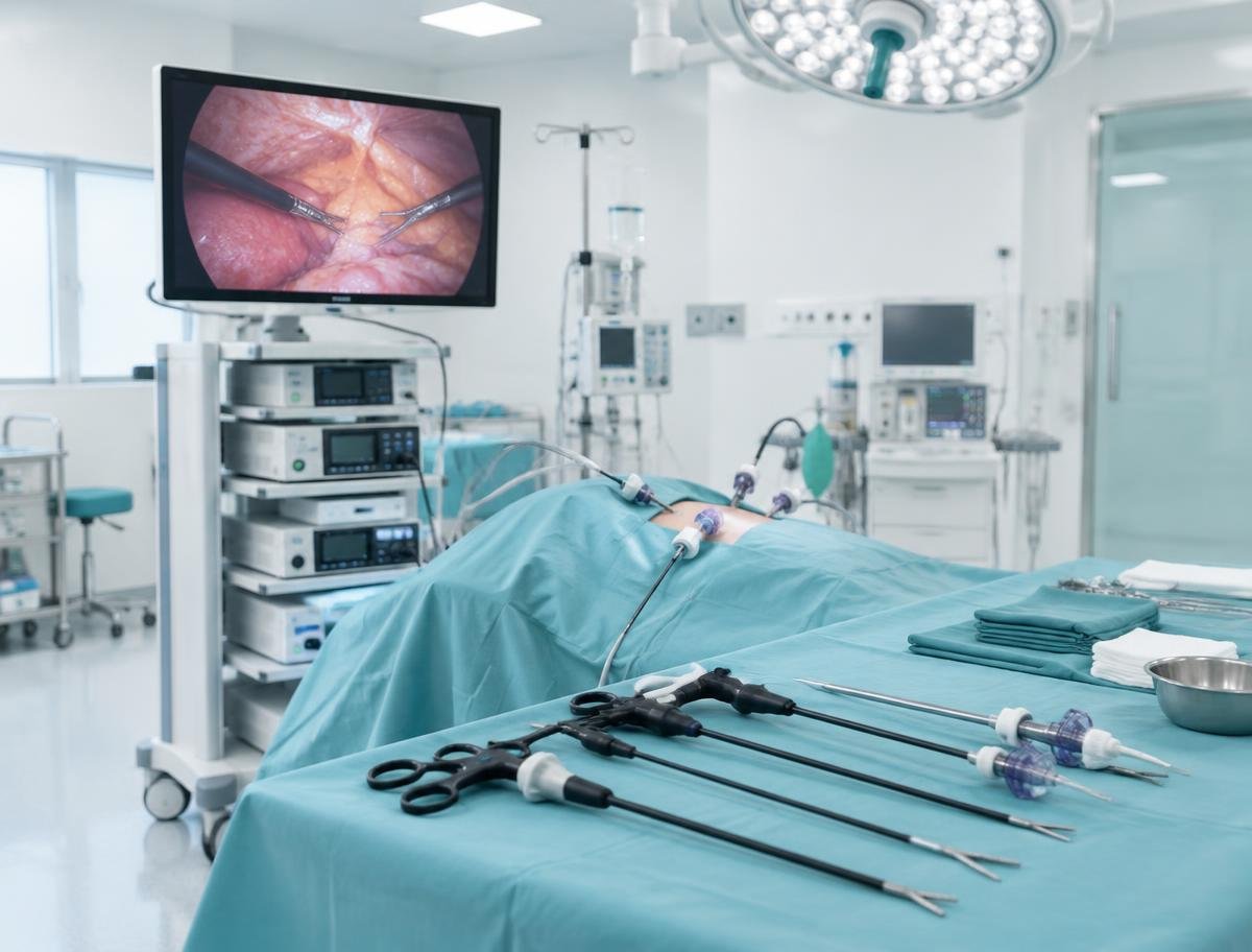
Understanding Hysteroscopy
No abdominal cuts are needed. The hysteroscope is guided through the vagina and cervix, and the uterus is gently expanded with fluid so its lining can be seen clearly on a screen.
A diagnostic hysteroscopy looks for problems; an operative hysteroscopy uses fine instruments through the same scope to remove polyps, small fibroids, adhesions or a septum.
Who may need this?
It may be considered in situations such as the following. Suitability is always decided after individual assessment.
- People with abnormal or heavy uterine bleeding.
- Those with suspected polyps, fibroids inside the cavity, adhesions or a uterine septum.
- Some people with recurrent miscarriage or repeated unsuccessful IVF.
- When scans show an abnormality of the uterine lining.
How does it work?
- 01
Assessment
Review of symptoms and scan findings to decide if hysteroscopy is appropriate.
- 02
Timing
Often planned after a period, before ovulation.
- 03
Procedure
The hysteroscope is inserted through the cervix; the cavity is examined and treated if required.
- 04
Discussion
Findings and any further care are explained afterwards.
Potential benefits
- Direct view of the uterine cavity.
- Many conditions can be treated in the same procedure.
- No abdominal incision.
Important considerations
- Possible risks include infection, bleeding and, rarely, injury to the uterus.
- Not every finding needs treatment; decisions are individual.
- Anaesthesia type depends on whether the procedure is diagnostic or operative.
Preparation and recovery
- Inform the clinic of any possibility of pregnancy.
- Follow fasting instructions if anaesthesia is planned.
- Bring previous ultrasound reports.
After the procedure
- Light cramping and spotting can last a few days.
- Most people return to normal activity within one to two days.
- Seek advice for heavy bleeding, fever or worsening pain.
This information is educational and does not replace an individual medical consultation. Outcomes vary and no treatment can guarantee a particular result.